
A microscope is a wonderful piece of technology, in that it allows us to look at our slide image wether its a red blood cell or the root of an onion in detail. "Micro" meaning small and "scope" meaning view. There are many different types of microscopes available, but the lab for this section was used with the Compound Light Microscope. Below is a quick description of the following pieces of the microscope: stage, focus knobs,iris,oculars, objectives.
Stage: The stage is where the slide is placed, with 2 clips to hold the slide into place. It moves up and down, but when the slide is in place ready to start viewing, make sure the stage is in highest position. Looking at the stage when moving it.
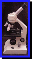
Focus knobs. The focus knobs are the coarse adjustment knob, which raises the image to a clear view when beginning the process. XY knobs move the slide up and down, left and right. The fine adjustment knob is used frequently, with slight turns, to get a clear/sharp view of the image. These knobs should be done while looking through the microscope.
Iris. The iris adjusts the light shining at the slide, while looking through the microscope. When changing objectives, its important to also adjust the iris.
Oculars. The oculars are located at the top of the microscope, also known as the eyepieces. These should be adjusted right away, an average distance is 64 for the right and left eyepiece, but should be adjusted to preference. Adjusting with both eyes open looking through the eye piece, 3/4 inches away.
Objectives. There are different objectives on the microscope, which basically are the magnification numbers. When starting to view a slide, the objective should be in 4x(shows the picture as a whole) and changed to higher magnification as preferred. When changing objectives, it's important to move the nose piece (where objectives are located) while looking at the microscope. Also when changing magnification to larger sizes: 10x, 40x ect. its important to readjust the iris.
Microscopes have come along way since it was first invented/used in 1595, by the Janssesn's. It basically consisted of a tube with a lens on each end of the tube. A couple other inventors that developed a more sophisticated microscope was: Hooke and Leeuwenhoek. Hooke is credited for discovering plant tissue under a microscope. Leeuwenhoek discovered bacteria in pond water and teeth. Other interesting individuals were the Egyptians, who used rock crystals shaped like lenses to view objects. The website shows many different and unique photos of microscopes used in previous years. With more experience and skills, microscopes developed into pieces of art.
 Janssesn's. It basically consisted of a tube with a lens on each end of the tube. A couple other inventors that developed a more sophisticated microscope was: Hooke and Leeuwenhoek. Hooke is credited for discovering plant tissue under a microscope. Leeuwenhoek discovered bacteria in pond water and teeth. Other interesting individuals were the Egyptians, who used rock crystals shaped like lenses to view objects. The website shows many different and unique photos of microscopes used in previous years. With more experience and skills, microscopes developed into pieces of art.
Microscopes prove to be useful pieces of equipment, since a lot of these objects can not be se en without the use of magnification! Now with the technology of today's world, there are far more sophisticated microscopes available. Research is very popular in today's society, which would be difficult to study without the use of the 3-D microscopes(a few examples): the Dissection Microscope and the Scanning/Transmission Electron Microscopes.
 en without the use of magnification! Now with the technology of today's world, there are far more sophisticated microscopes available. Research is very popular in today's society, which would be difficult to study without the use of the 3-D microscopes(a few examples): the Dissection Microscope and the Scanning/Transmission Electron Microscopes.
No comments:
Post a Comment