MUSCLE CONTRACTION- LAB
As you are bending down to pick up a penny off of the ground, a process occurs in your limbs and tissues in order for this movement to take place. This lab project will hopefully give you a better understanding of how a muscle moves; my example is of a knee joint. Skeletal muscle is one the the 3 muscle tissues in the body, which are needed in order for a contraction to occur. "Skeletal muscle tissue moves the body by pulling on bones of the skeleton" (Martini 284). Within the skeletal muscle tissue is a single muscle fiber. My lab project shows in detail the start of a contraction, which occurs when an electric impulses travels down the length of a muscle fiber. Before it even gets to the muscle fiber, a motor neuron is the carrier of the information that brings the impulses away from the CNS to the muscle fiber. The axon is a snakelike figure that extends off of the motor neuron, responsible for conducting nerve impulses. I will also be describing the process of action potential, which is a nerve impulse traveling down the axon. At this point by reviewing the pictures will hopefully give a better understanding of how a muscle begins a contraction. There are 6 functions of a skeletal muscle: Movement, maintain body position and posture, support soft tissues, protect particles that enter and exit, maintain body temperature, and store nutrients. These functions are all necessary in order to provide a healthy movable muscle. 


Above is a picture of a movable limb: a femur bone, tibia bone and between is the synovial joint. The middle picture is the same limb, but showing the Sartorium muscle, which helps with flexion of the knee and hip and also with rotation of the hip. There are several classifications of synovial joints, which basically allows for different types of range of motion. The knee joint can be described as a hinge joint, which allows motion similar to opening and closing of a door. The picture to the far right is a picture off of a website, but in more detail of the same limb I used.

Above is a picture of a motor neuron(green foam) the white strings off of the neuron are dendrites, the skinny white string is an axon, the pink beads represent the mylein sheath which is covered by Schwann cells, and the twizzler is a muscle fiber. As mentioned above the motor neuron carries nerve impulses away from the CNS to a muscle fiber. Messages are received from other neurons at the dendrites(strings off of the motor neuron). These messages are then passed onto the axon(skinny white string), in a form known as an action potential. At the end of each axon are synaptic terminals( a synapse is also a part), which is involved in the communication method to other cells. This picture basically shows how the neurons are moving towards a muscle cell, sending messages down the axon in form known as action potential getting ready to start a contraction.

This picture shows where the action potential occurs. It moves along the axon, stimulated from the electrical impulses. At first I used a white string for my axon, but for vision reasons I switched to a twizzler.


The next process is that of nerve impulses. Basically it is information that is moving along the axon, in the form of action potential, the electrical impulses are known as nerve impulses. There are 2 types of potentials: Resting(axon has no impulse movement, -65mV) and Action("rapid change in polarity across an axonal membrane as the nerve impulse occurs") (Mader 250). The pictures above are both Action potential. The first one is when the Sodium gates open(green balls are sodium) and Sodium enters the axon. The Potassium gate (blue structure) remains closed at this time. During this process, depolarization occurs, which is caused from the axon becoming more positive with the entrance of sodium. "The potential changes from -65mV to +40mv" (Mader 250). The next picture shows reporization: Potassium ions move out of the axon making the inside negative again. During this phase, Sodium gates are closed and Potassium gates swing open.

This picture is inside of the muscle fiber, showing the T tubule(black strings) and the sarcoplasmic reticulum (pink thread). The plasma membrane of a muscle fiber is known as the sarcolemma. T tubules are networks inside of the muscle fiber that carry electrical impulses to the cell. One of the important parts of the sarcoplasmic reticulum is that it is a storage unit for Calcium ions. The spider web structure formed from the T tubules and sarcoplasmic reticulum hold together hundreds of myofibrils. Inside the myofibril are many sacromeres. These structures are bundled with proteins: Thin (actin) and thick(myosin); these protein molecules are responsible for the muscle fiber to contract.

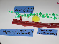
This picture shows the 2 different filaments I just mentioned. Note the little flaps on the Myosin structure(twizzler) these are called myosin heads, later I will show the interaction of the head connecting to the Actin, these are known as cross-bridges. The actin is intertwined with 2 strands: tropomyosin(pink string) and troponin(large green beads).
The start of the contraction begins when the neurotransmitter acetylcholine(ACh) is released and binds to a messenger in the sarcolemma(plasma membrane of the muscle fiber). This impulse travels down the network of T tubules and ends intertwining sarcoplasmic retiuclum. Calcium ions are released which leads to a sacromere contraction. The picture to the far right shows the release of calcium ions(yellow balls) which attach to actin filaments- on the troponin protein thread(large green beads).
Once the Calcium ions are attached to the Troponin, this causes the tropomyosin(pink string) to shift away, making room for mysoin to bind to actin. The picture above shows this process. The head of the mysoin attached to the actin filament. At this point ATP supplies the needed energy for a contraction to occur. The cross-bridges as mentioned above are a part of this process. Contraction beings when this cycle of cross-bridges binding, pivoting, and detachment occurs repeatedly. "The muscle fiber contracts as the sarcomeres, within the myofibrils, shorten" (Mader 232). Its difficult to show how sliding filaments work, but the actin filament slides toward the center of the sacromere when the head of the myosin pivots to the base of the actin filament. "When muscle cells contract, they create tension and pull on the attached tendons" (Martini 321).

In conclusion, our bodies are made of many cells, tissues, and muscles that help keep us functioning every second. This lab was definitely a piece of work, but it gave me a strong understanding of how a muscle contraction occurs. The skeletal muscle contains many muscle fibers packed with myofibrils. The myofibrils consist of sacromeres which are composed of thick(myosin) and thin(actin) filaments. When a sacromere shortens in length and actins slide past myosin, a muscle contraction occurs. The picture to the right is a quick outline of what I described throughout this lab. It's amazing how all of these processes I described occur in order for a muscle to move. Hopefully my write-up was helpful in showing how a neuron moves through the body just to allow you to bend a pick up the penny off of the ground.
Works cited:
picture of knee-muscles
Mader, Sylvia S. Human Biology. Boston: McGrawHill companies, Inc. 2008.
quotesMartini, Fredric H. Fundamentals of Anatomy and Physiology. San Francisco: Pearson Education, Inc. 2006.
quotes


1 comment:
Amanda,
Stellar, really perfect work on this unit. I really like your limb model and appreciated your essay perspective. Also, nice organized compendium reviews. Keep it up and only one unit to go!
LF
Post a Comment